Home
Uncategories
Cross Section Of A Bone / Cross Section Bone Human High Resolution Stock Photography And Images Alamy / To the left is muscle tissue, and to the right is bone marrow.
Cross Section Of A Bone / Cross Section Bone Human High Resolution Stock Photography And Images Alamy / To the left is muscle tissue, and to the right is bone marrow.
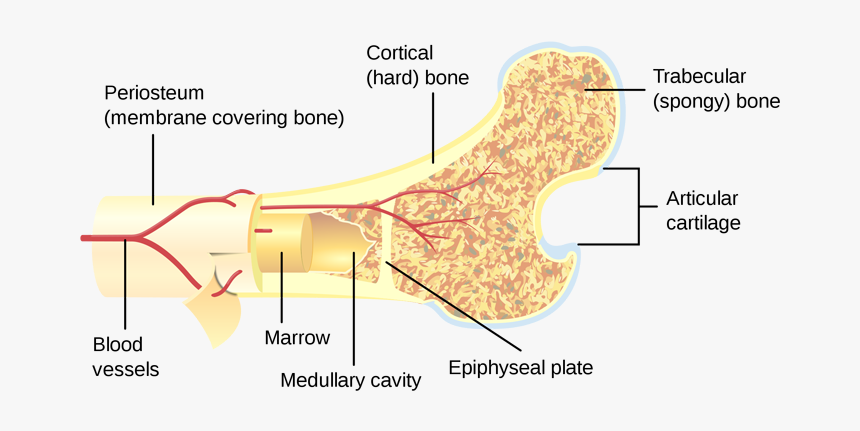
Cross Section Of A Bone / Cross Section Bone Human High Resolution Stock Photography And Images Alamy / To the left is muscle tissue, and to the right is bone marrow.. Internal structure of a human long bone, with a magnified cross section of the interior. Cross section of mandible at first molar region showing cortical and spongy bone basic concepts in osteogenesis bone is a dynamic biological tissue, composed of various metabolically active cells that are integrated into a rigid framework. Thus, the lamellar pattern as well as the lacunae size differ between trabecular and cortical bone. Compact bone is the outer layer and the spongy bone forms the inner layer. Smartdraw includes 1000s of professional healthcare and anatomy chart templates that you can modify and make your own.
The remainder is cancellous bone, which has a spongelike appearance with numerous large spaces and is found in the. Bone markings the surface features of bones vary considerably, depending on the function and location in the body. Compact bone is the outer layer and the spongy bone forms the inner layer. If i can teach you one thing about how to draw the back of a person, it's that it's absolutely crucial to understand the position of the scapula bones (shoulder in this tutorial, we will go over the bones and major muscle groups you will need to know to draw the. The upper (biting) surfaces of the tooth are at top, with the lower sections (bottom) embedded in the gums and jaw bone (not shown).
Cross Section Of A Bone Hd Png Download Kindpng from www.kindpng.com An outer 'fibrous layer' containing mainly fibroblasts, and an inner 'cambium layer' containing progenitor cells. This slide contained a cross section of a very small bone, and you are looking at the entire thickness of the shaft of the bone. I don't find it enhances the image. Compact bone, spongy bone, and bone marrow. Table 1 describes the bone markings, which are illustrated in (figure 4). Browse 53 bone marrow cross section stock photos and images available, or search for bone cross section or bone cells to find more great stock photos and pictures. Now that you know what bones do, let's take a look at what they're made of and their anatomy. Start studying cross section of long bone.
Compact bone is the outer layer and the spongy bone forms the inner layer.
The remainder is cancellous bone, which has a spongelike appearance with numerous large spaces and is found in the. Browse 53 bone marrow cross section stock photos and images available, or search for bone cross section or bone cells to find more great stock photos and pictures. Bone matrix and cells bone matrix osseous tissue is a connective tissue and like all connective tissues contains relatively few cells and large amounts of extracellular matrix. An outer 'fibrous layer' containing mainly fibroblasts, and an inner 'cambium layer' containing progenitor cells. Cross‐sectional area is derived from the integral of the bone mass profile across the narrow region. Cross section of a human bone showing bone marrow, spongy bone and blood vessels. Learn vocabulary, terms, and more with flashcards, games, and other study tools. Start studying cross section of bone. The cortical bone equivalent area of the cross‐section of the region of interest (femoral neck or shaft), with all soft tissue voids (trabecular and cellular spaces) eliminated (cm 2). Compact bone, also called cortical bone, dense bone in which the bony matrix is solidly filled with organic ground substance and inorganic salts, leaving only tiny spaces (lacunae) that contain the osteocytes, or bone cells.compact bone makes up 80 percent of the human skeleton; There are trabeculae in spongy bone which gives its sponge like appearance. Each bone in your body is made up of three main types of bone material: Cross section of mandible at first molar region showing cortical and spongy bone basic concepts in osteogenesis bone is a dynamic biological tissue, composed of various metabolically active cells that are integrated into a rigid framework.
The remainder is cancellous bone, which has a spongelike appearance with numerous large spaces and is found in the. Why is the marrow red? Bone markings the surface features of bones vary considerably, depending on the function and location in the body. It consists of two layers; While it is not as hard as compact bone, spongy bone plays an important role of protecting the marrow where blood cells are produced.
Cross Sectional Anatomy Kenhub from thumbor.kenhub.com Now that you know what bones do, let's take a look at what they're made of and their anatomy. Cross section of a human bone showing bone marrow, spongy bone and blood vessels. Compact bone, also called cortical bone, dense bone in which the bony matrix is solidly filled with organic ground substance and inorganic salts, leaving only tiny spaces (lacunae) that contain the osteocytes, or bone cells.compact bone makes up 80 percent of the human skeleton; There are three general classes of bone. Internal structure of a human long bone, with a magnified cross section of the interior. Start studying cross section of long bone. While it is not as hard as compact bone, spongy bone plays an important role of protecting the marrow where blood cells are produced. Human back muscles and bones 12 photos of the human back muscles and bones human back muscles and bones, bone, human back muscles and bones
There are three general classes of bone.
An outer 'fibrous layer' containing mainly fibroblasts, and an inner 'cambium layer' containing progenitor cells. Human back muscles and bones 12 photos of the human back muscles and bones human back muscles and bones, bone, human back muscles and bones Learn vocabulary, terms, and more with flashcards, games, and other study tools. It consists of two layers; Related posts of cross section of a long bone bone test anatomy and physiology. Cross‐sectional area is derived from the integral of the bone mass profile across the narrow region. The large dark spots are passages for blood vessels and nerves. The compact bone is made up of osteon. 100x first focus in the compact decalcified bone (cb) on the left part of the image, you can see small dots, which are. The surface features of bones vary considerably, depending on the function and location in the body. Each bone in your body is made up of three main types of bone material: Two types of bone tissues in cross section of a long bone : Compact bone, also called cortical bone, dense bone in which the bony matrix is solidly filled with organic ground substance and inorganic salts, leaving only tiny spaces (lacunae) that contain the osteocytes, or bone cells.compact bone makes up 80 percent of the human skeleton;
It consists of two layers; As the names suggest compact bone looks compact and the spongy bone looks like sponges. Browse 4,275 bone cross section stock photos and images available, or search for human bone cross section to find more great stock photos and pictures. 100x first focus in the compact decalcified bone (cb) on the left part of the image, you can see small dots, which are. Compact bone, also called cortical bone, dense bone in which the bony matrix is solidly filled with organic ground substance and inorganic salts, leaving only tiny spaces (lacunae) that contain the osteocytes, or bone cells.compact bone makes up 80 percent of the human skeleton;
Q Wciggpo9h1am from images.freeart.com Smartdraw includes 1000s of professional healthcare and anatomy chart templates that you can modify and make your own. Lamellar bone makes up the compact or cortical bone in the skeleton, such as the long bones of the legs and arms. After a fracture, woven bone forms initially and is gradually replaced by lamellar bone during a process known as bony substitution. To the left is muscle tissue, and to the right is bone marrow. This slide contained a cross section of a very small bone, and you are looking at the entire thickness of the shaft of the bone. The compact bone is made up of osteon. Browse 53 bone marrow cross section stock photos and images available, or search for bone cross section or bone cells to find more great stock photos and pictures. Compact bone, spongy bone, and bone marrow.
Marrow in the shaft of long bones is typically yellow, with red marrow in the head through the cancellous bone.
Two types of bone tissues in cross section of a long bone : The remainder is cancellous bone, which has a spongelike appearance with numerous large spaces and is found in the. Browse 53 bone marrow cross section stock photos and images available, or search for bone cross section or bone cells to find more great stock photos and pictures. Table 1 describes the bone markings, which are illustrated in (figure 4). Start studying cross section of long bone. Related posts of cross section of a long bone bone test anatomy and physiology. Learn vocabulary, terms, and more with flashcards, games, and other study tools. Thus, the lamellar pattern as well as the lacunae size differ between trabecular and cortical bone. The upper (biting) surfaces of the tooth are at top, with the lower sections (bottom) embedded in the gums and jaw bone (not shown). Start studying cross section of bone. Human back muscles and bones 12 photos of the human back muscles and bones human back muscles and bones, bone, human back muscles and bones To the left is muscle tissue, and to the right is bone marrow. 100x first focus in the compact decalcified bone (cb) on the left part of the image, you can see small dots, which are.

0 Comments:
Posting Komentar